09 Feb 2024
HPLC Strategies for the Characterisation Of AAV Gene Therapy Vectors
Adeno Associated Viruses (AAVs) are members of the Parvovirus family of animal viruses. Parvoviruses virions are small compared to most viruses, at 23-28 nanometers in diameter. AAVs can infect human and primate cells and have proven a promising vector in gene therapy. Critically, AAVs exhibit a low genotoxicity profile in humans, integrate in a stable and persistent manner and the AAV genome remains inactive. These properties make AAVs suitable viral vectors for the introduction of intact genes into human cells in a stable and sufficiently targeted manner, without causing disease or stimulating a severe immune response. In addition, their genome structure is less complex, which simplifies experimental manipulation. A wide range of AAV variants (serotypes) are known today. Their envelope proteins have unique surface structures; accordingly, they differ in the choice of their target cells (tropism). Since a given vector genome can easily be packaged in the envelope of any AAV serotype, there are few cell types which would not be amenable to the introduction of AAV vector DNA. This versatility and their natural simplicity have made AAVs the most important vector in the advancement of gene therapies. [1]

AAV gene therapies and any precursor starting materials must be well characterised to fully understand the relationship between product quality attributes, manufacturing processes, patient safety and efficacy.
The two basic components of a serotherapeutic reagent are the viral capsid (shell) and the nucleic acid material within the capsid, that will be delivered by the vector to the target cell. The AAV capsid is composed of three viral proteins (VPs): VP1 (≈90 kDa), VP2 (≈68 kDa), VP3 (≈62 kDa). The molar ratio of the single proteins is estimated to be 1:1:10. Altered ratios, for example due to degradation, may change the vectors potency and carry an increased risk of immunogenicity. Therefore, the quantity each of these proteins needs to be closely controlled during product development to ensure drug safety. [2]
A variety of non-chromatographic analytical methods exist (e.g., qPCR, ddPCR, NGS, Western blot, ELISA, Flow cytometry, dynamic light scattering-DLS and AUC, EM, SDS-page gels, and differential scanning fluorimetry-DSF) that are routinely used in manufacturing and development testing. However, many of these approaches lack accuracy and precision and require extensive method development or re-qualification whenever the matrix of the sample is changed due to limited specificity. [3]
Several different HPLC separation techniques provide higher resolving power and increase accuracy, precision, and reproducibility in comparison with non-chromatographic analysis. These approaches are today favoured for both in process testing and development.
Within the United States Pharmacopeia (USP) guidelines – Analytical Methods for drug substance and drug product, HPLC methods are recommended for the following Critical Quality-Attributes:
- Identity: Viral Capsid (Reversed Phase HPLC-Mass Spectrometry (RP-LC/MS))
- Quantity: Total viral capsid particles (Size Exclusion Chromatography-Multi-Angle Light Scattering (SEC-MALS), Anion Exchange Chromatography (AEX-HPLC)
- Purity: Capsid content – empty vs full (Anion Exchange Chromatography (AEX-HPLC)) [5]
Viral Capsid Identity
Viral Protein (VP) identification and quantification can be performed by Reversed Phase HPLC linked to Mass Spectrometric detection. However, this can be a challenging separation as all three proteins are spliced from the same gene and exhibit high sequence homology. This is problematic because coelution may compromise signal intensity and mass accuracy, in addition to precluding accurate quantitation of capsid stoichiometry, which is an important determinant of AAV infectivity [4]. Choosing the correct HPLC column that gives the optimum selectivity, high efficiency and low nonspecific interactions is paramount if resolution is to be achieved.
In this example the separation is performed using a YMC-Triart Bio C4 column. With its wide pores of 300 Å and less hydrophobic butyl surface modification, the YMC-Triart Bio C4 column enables the separation of large biomolecules of up to 150 kDA. Achieving resolution between the three VPs may not be possible on all C4 columns because retention factors are close. YMC Triart-C4 exhibits an optimised selectivity compared to other C4 columns and successful separation can be achieved. YMC-Accura Triart-C4 uses bioinert hardware, which stops protein interaction that often occurs in typical stainless steel column hardware and improves the quality of the separation even further.

Figure 1. Comparison of VPs separation by RP-LC using the bioinert YMC-Accura Triart Bio C4, YMC-Triart C4 (standard hardware) and another C4 column with stainless stell hardware.
| Columns: | YMC-Accura Triart Bio C4 (1.9 µm, 30nm) 150 x 2.1mm ID (bioinert hardware) YMC-Bio C4 (1.9 µm, 30nm) 150 x 2.1mm ID (standard hardware) Other C4 column (1.7 µm, 30nm) 150 x 2.1 ID |
| Part Nos: | TA30SP9-15Q1PTC TA30SP9-15Q1PT |
| Eluent: | A. water + 0.1% difluoroacetic acid B. acetonitrile + 0.1% difluoroacetic acid |
| Gradient: | 20-32%B (0-1 min), 32-36%B (1-16 min), 36-80%B (16-20 min) |
| Flow Rate: | 0.2mL/min |
| Detection: | UV at 280 nm ESI-MS (positive ion mode) |
| Temperature: | 80°C |
| Injection: | 50µL |
| Sample: | Denatured AAV2 |
YMC-Accura Triart-C4 is an excellent choice for the analysis of AAV VPs since it offers the following advantages over other C4 columns.
- Improved separation
- Better peak shapes
- Reduced retention time
Different stationary phase selectivity can be useful in developing methods for intact AAV VP identification in different serotypes. In this example a rapid identification LC/MS method is developed using Zorbax RRHD 300 1.8um columns with three different selectivities 300SB-C18, 300-Diphenyl and 300SB-C3. For AAV-2 300SB-C18 offers superior separation of VP-1 with greatly reduced interference by VP3, permitting reliable and accurate quantitation (Figure 2 and Figure 3)
Featured Product

Figure 2. Method development with AAV-2. Fluorescence chromatograms depicting separation using (A) Agilent ZORBAX RRHD 300Å SB-C3, (B) SB-Diphenyl, and (C) SB‑C18 columns. Note the superior chromatographic resolution of SB‑C18. (D to F) Deconvoluted mass spectra of chromatographic peaks showing the coelution of VP1 and VP3.

Figure 3. LC/MS of denatured AAV-2 capsid proteins using the optimized method on an Agilent ZORBAX RRHD 300Å SB-C18 column. (A) Fluorescence chromatogram depicting the three capsid proteins. (B) Total ion current. (C to E) Deconvoluted mass spectra of the three capsid proteins, with the relevant mass peaks marked by asterisks.
To assess the generalizability of this LC/MS method to the analysis of other AAV serotypes, AAV-7, 7m8, DJ, 9, rh10, and Anc80 were analyzed using the optimized SB-C18 method described above. AAV-7, 7m8 and DJ separated well using this method (Figures 4A to 4C), but AAV-9, rh10, and Anc80 separated as only two peaks, with VP1 and VP3 coeluting as single peaks. Agilent diphenyl stationary phases have altered selectivity compared to alkyl chain stationary phases such as C3, C8, and C18. The phenyl groups permit separation based on π‑π interactions with additional affinity for aromatic amino acids and double bonds. An optimized SB‑Diphenyl method was developed to achieve successful separation of AAV-9, rh10, and Anc80 (Figures 4D to 4F)

Figure 4. LC/MS of denatured AAV capsid proteins of six different serotypes on (A to C) Agilent ZORBAX RRHD 300 SB-C18, and (D to F) Agilent SB-Diphenyl columns.
Aggregation and Fragment Analysis
Aggregate studies are performed using size exclusion chromatography (SEC) coupled to multi-angle light scattering (MALS) detection. AAVs although small in comparison with other viruses are still large molecular complexes consisting of approximately 60 copies of capsid protein(s) encapsulating a single stranded DNA genome. Individual AAV virions may exceed 5 MDa and are approximately 250 Å in size. AAV aggregates are therefore challenging to resolve using standard SEC columns, which typically have pore sizes ≤300 Å. Conventional wisdom indicates that an SEC pore size at least three times larger than the molecule of interest should be chosen, which would indicate a pore size ≥750 Å.
In this example AAV 2 and 9 serotypes are analysed using an Agilent Bio SEC-5 column with 1,000 Å pore size. Agilent Bio SEC-5 columns are packed with 5 μm silica particles coated with a proprietary hydrophilic layer for efficiency and stability, and are thus well suited for analysis of large, complex biological molecules such as viral particles.
Featured Product

Figure 5. Fluorescence chromatograms of Agilent AdvanceBio SEC 300Å Protein Standard and unstressed AAV-9 and AAV-2. Note the protein standard also contains insulin, which is not shown because it contains no tryptophan amino acids and is therefore nonfluorescent under the chosen excitation and emission settings.

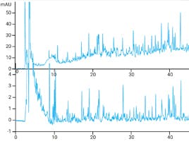
Figure 6. Fluorescence chromatograms of unstressed and stressed AAV-9.
As expected, both AAV-2 and AAV-9 show elution times between thyroglobulin monomers and their aggregates (Figure 5) Unstressed AAV-2 and AAV-9 had similar purity values of 98.7% and 98.8% respectively, each containing 1.2 to 1.3% aggregates. In contrast, stressed AAV-9 showed extensive formation of aggregates and substantial levels of degradation. Stressed AAV-9 was 83.6% pure, containing 7.5% aggregates and 8.8% fragments. Notably, the total peak area of stressed samples was only approximately 60% that of unstressed samples, possibly indicating the formation of insoluble aggregates (Figure 6).
Other columns that can be successfully used for aggregate and fragment analysis include;
- TSKgel G6000PWxl
- SEPAX SRT SEC-750 A
- Thermo Scientifc Acclaim SEC-1000
Capsid Purity-Full Vs Empty
A key component of a successful AAV workflow is obtaining a high overall yield of full viral capsids, along with efficient removal of empty capsids, which are essentially an impurity. Ion Exchange chromatography utilizes small differences in the average isoelectric point (pI) between full capsids (pI 5.9) and empty capsids (pI 6.3). This can be achieved by using anion exchange chromatography (AEX). [5]
In this example a strong anion exchange stationary phase YMC BioPro IEX QF is used for the separation. YMC BioPro IEX QF columns are based on non-porous hydrophilic polymer beads with low nonspecific adsorption. They also show higher binding capacity and higher recovery of biomolecules compared to conventional ion exchange columns. The completely spherical, monodisperse packing materials provide excellent peak shape.
Featured product
Figure 7. Empty/Full Capsid separation by AEX-HPLC using a YMC BioPro IEX QF column.
| Column: | BioPro IEX QF (5 µm) 30 x 4.6mm ID |
| Eluent: | A. 20mM Bis-tris propane-HCl (pH 9.0) B. 20mM Bis-tris propane-HCl containing 0.5 M (CH3)4NCl (pH 9.0) 5%B (0-0.2 min), 20-45%B (0.2-10 min) |
| Flow Rate: | 0.5mL/min |
| Detection: | FLS at Ex.280 nm, Em. 348 nm |
| Temperature: | 25°C |
| Injection: | 2µL |
| Sample: | AAV2 (2.59 x 10¹² vg/mL) |
Other columns that can be successfully used for capsid purity testing include;
- Agilent Bio SAX
- Thermo Scientifc ProPac 3R SAX
Since AAV is relatively small for a virus (20-25 nm) this makes it amenable to separation using both traditional and monolithic stationary phase support materials. Monolithic columns such as Agilent Bio-Monolith QA are sometimes preferred when dealing with very large viruses and biomolecules as there is less risk of blockage, higher flow rates may be used, and fewer shear forces which may cause mechanical disruption of the analyte.
Summary
This article has highlighted some of the analytical HPLC techniques used determining Critical Quality Attributes in AAV Gene Therapy Vectors. AAV testing poses challenges since the parameters which define cell and gene therapy product safety are still emerging. Choosing the correct HPLC column for your application is paramount.
As your chromatography partner, Element Lab Solutions can provide support with all aspects of HPLC method development and optimisation. We have a wide selection of high-quality analytical columns from a range of manufacturers, suitable for a broad range of biomolecule applications. For advice on column selection, method development or troubleshooting of AAV analysis, our in-house technical team are always on hand to provide expert support.
To learn about the techniques described in more detail and other chromatographic strategies for biomolecule analysis head over to CHROMacademy, where there are a huge number of resources on these topics.
References
2. https://www.sciencedirect.com/science/article/pii/S0165993623001759
3. https://www.ncbi.nlm.nih.gov/pmc/articles/PMC8780015/
4. https://www.ncbi.nlm.nih.gov/pmc/articles/PMC9910337/
6. Bosma, B. et al. Optimization of Viral Protein Ratios for Production of RAAV Serotype 5 in the Baculovirus System. Gene Therapy 2018, 25(6), 415–424.
Related articles